The 2.2 Å structure of a full-Length catalytically active hammerhead Ribozyme
Monika Martick and William G. Scott
Copyright, 2006
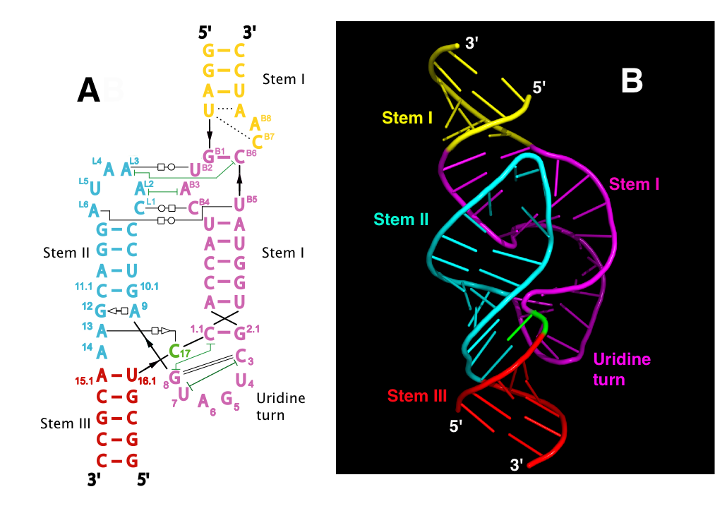
The secondary and tertiary structure are shown with various tertiary contacts indicated in the diagram
on the left. The color-coding of the sequence matches that of the structure.

The cleavage-site nucleotide is now positioned for in-line attack. G-12 is positioned for general base catalysis, and G-8 may assist as a general acid. The non-bridging phosphate oxygens of the A-9 and scissile phopshate are 4.1 Å apart, and could potentially bind a divalent metal ion, although none are seen to be bound in the crystal structure.
- High resolution figures and other supplemental stuff (including a movie).
- Martick and Scott: Tertiary Contacts Distant from the Active Site Prime a Ribozyme for Catalysis. Cell, 126:309-320 (2006). (Local pdf copy).
- Nelson and Uhlenbeck: When to Believe What You See. Molecular Cell, 23:447-450 (2006).
- Eric Westhof: A tale in molecular recognition: the hammerhead ribozyme. J. Molecular Recognition, 20:1-3 (2006).
- Przybilski and Hammann: The Hammerhead Ribozyme Structure Brought in Line. ChemBioChem 7: 1641-1644 (2006).
- View interactively via Jmol